The central goal of the Hébert Lab is to reconstruct neocortical tissue in such a way that the activity of this new tissue can encode useful behavior to the animal. One strategy for approaching this goal is by combining a normal complement of neocortical precursor cell types for transplantation, including neuronal, glial, and vascular subtypes, and arranging the precursors in a laminar cytoarchitecture that resembles the normal developing neocortex.
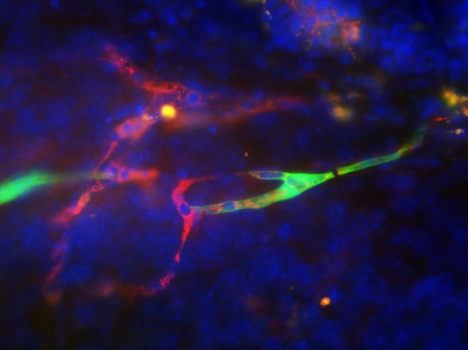
Live imaging of a human neocortical neuron 2 days after transplantation into the adult mouse neocortex showing a growth cone exiting the graft and finding its way.
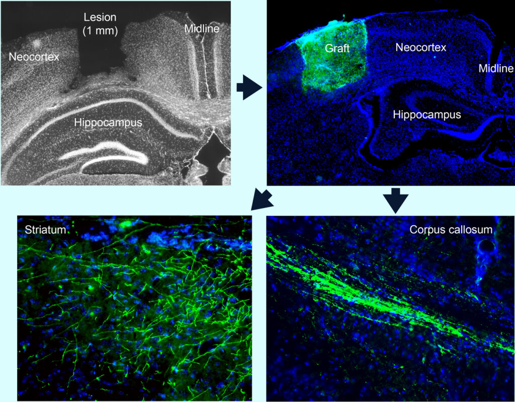
A key aspect to obtaining functional cortical tissue is cytoarchitecture. To minimize experimental variability, we can standardize the size of neocortical lesions with the aim of optimizing the deposition of a structured, cellularized, and vascularized bioscaffold. Within 2 weeks after transplantation, the graft is well vascularized (see below) and its neurons project and connect to normal targets, namely to the contralateral cerebral hemisphere across the corpus callosum and also down to the striatum.
When vascular endothelial cells are included in the transplant cell population, they form vessels (green) and integrate with the host vasculature to circulate blood.
Live imaging of human vascular endothelial cells 30 days after transplantation (with human neuronal stem cells), showing extensive vessel formation within the graft.
We can visualize and measure blood circulation in live animals by labeling a small fraction of erythrocytes with DiO (rapidly moving green dots).
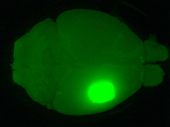
A graft whose cells are labeled with GFP (green fluorescent protein) 3 months post-transplantation, as part of a study to determine how young grafts affect the brains of old mice (>24 months), and how old brains affect young grafts.
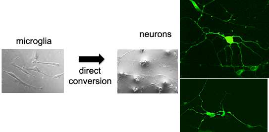
Another goal of the Hébert Lab is to use microglia as a vehicle for the long-term and widespread delivery of biologics or cells to the neocortex.
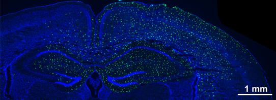
The lab has developed a protocol that allows for a single injection of engineered microglia (small green dots) to distribute and replace their endogenous counterparts throughout large areas of cortex.
A potential application for microglia dispersion in the cortex that is being explored is the replacement of neurons in age-related degeneration via direct conversion of engineered microglia to neocortical neurons.